Cerebral angiography
Imaging technique for brain blood vessels, pioneered in 1927.

Cerevisae · CC BY-SA 4.0
Cerebral angiography is a medical imaging technique that shows blood vessels inside and around the brain, helping doctors find problems like arteriovenous malformations and aneurysms. Portuguese neurologist Egas Moniz first developed it in 1927 at the Hospital de Santa Marta in Lisbon, and he used thorotrast as a contrast agent, though the substance was developed by others.
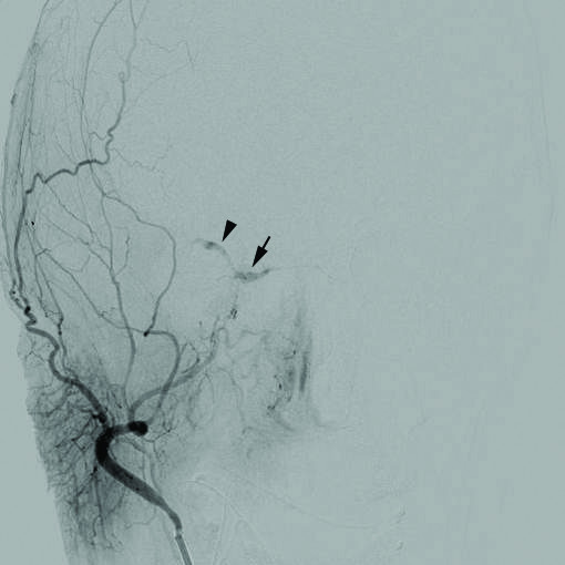
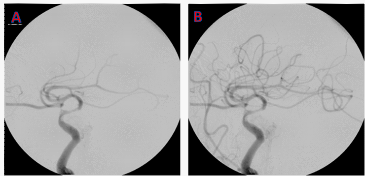
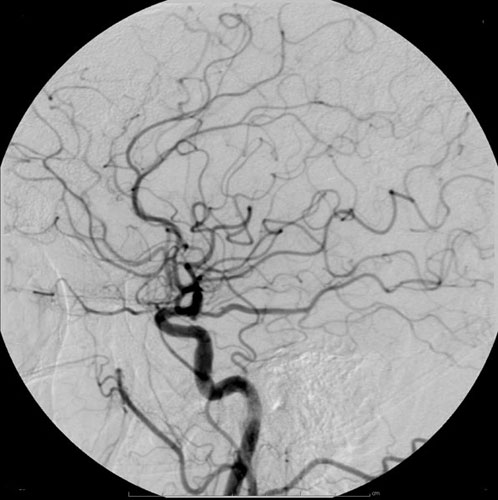
The procedure usually involves inserting a catheter into a large artery, often the femoral artery, and guiding it through the circulatory system to the carotid artery. A contrast dye is then injected, and a series of X-ray images are taken as the dye moves through the brain's arteries, followed by another set as it reaches the veins. For some cases, cerebral angiography can produce clearer images than less invasive methods like CT angiography or MR angiography. It also allows doctors to perform certain treatments right away based on what they see. In recent decades, the technique has taken on a more therapeutic role due to advances in endovascular treatments. For example, embolization—a minimally invasive procedure—has become increasingly important in treating cerebral arteriovenous malformations, often making later surgery or radiosurgery easier. If an aneurysm is found, doctors can insert metal coils through the catheter directly into the aneurysm; over time, these coils encourage connective tissue to form, strengthening the vessel wall.
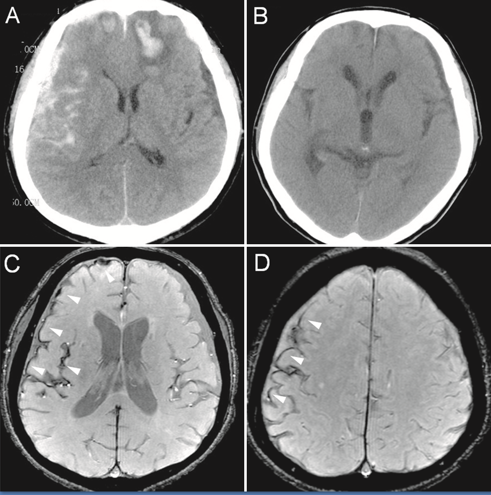
Before modern imaging tools like MRI and CT became available in the mid-1970s, cerebral angiography was often used indirectly to find certain lesions or hematomas by looking at how they displaced blood vessels. That use is now outdated, since non-invasive methods can directly image many intracranial problems. Still, cerebral angiography remains widely used to evaluate various vascular conditions inside the skull.
Cerebral angiography is used for diagnosis, but treatment can follow in the same session. It images both intracranial (inside the head) and extracranial (outside the head) diseases. Intracranial conditions include non-traumatic subarachnoid hemorrhage, non-traumatic intracerebral hemorrhage, intracranial aneurysm, stroke, cerebral vasospasm, cerebral arteriovenous malformation (for grading and planning intervention), dural arteriovenous fistula, embolization of brain tumors like meningioma or cavernous sinus hemangioma, the Wada test, and me
- pioneer
- Egas Moniz
- year_pioneered
- 1927
- field
- Neurology, Radiology
- nationality
- Portuguese
- known_for
- Pioneering cerebral angiography and using thorotrast as a contrast agent for the procedure
Lore & Background
Cerebral angiography is performed by inserting a catheter into a large artery, such as the femoral artery, and threading it to the carotid artery, where a contrast agent is injected. A series of radiographs are taken as the contrast agent spreads through the brain's arterial system, then a second series as it reaches the venous system. The procedure allows for both diagnosis and immediate treatment, such as embolization of arteriovenous malformations or coiling of aneurysms.
Reader's Guide
Cerebral angiography remains significant for its high-resolution imaging of blood vessel lumens and vasculature, providing better images than less invasive methods like computed tomography angiography and magnetic resonance angiography for some applications. It is the standard for detecting intracranial aneurysms and evaluating the feasibility of endovascular coiling. Although prior to the advent of modern neuroimaging techniques in the mid-1970s it was used indirectly to infer lesions and hematomas, that use is now obsolete. However, it is still widely used for evaluating various intracranial and extracranial vascular pathologies, and its therapeutic role has grown with endovascular techniques such as embolization and coiling.
Did You Know?
- Cerebral angiography was pioneered in 1927 by Portuguese neurologist Egas Moniz at the Hospital de Santa Marta in Lisbon.
- The procedure can be used to treat aneurysms by introducing metal coils through the catheter to encourage connective tissue formation.
- Digital subtraction angiography
Origins & Pioneering Work
Cerebral angiography traces its roots to 1927, when Portuguese neurologist Egas Moniz, working at the University of Lisbon, first demonstrated the technique. Moniz was instrumental not only in establishing the procedure but also in helping develop thorotrast, a contrast medium used to make the brain's vasculature visible under X-ray. This early work laid the groundwork for what would become an essential tool in neurovascular medicine. The procedure's core principle—introducing a contrast agent into the cerebral arterial system and capturing a sequence of radiographs as the dye travels through both arterial and venous networks—has remained fundamentally unchanged since Moniz's era. What has evolved dramatically is the precision of the equipment, the range of conditions it can address, and the therapeutic interventions that can now be performed through the same catheter access. Moniz's contribution established the paradigm of using vascular imaging to detect abnormalities such as arteriovenous malformations and aneurysms, a role that cerebral angiography continues to fulfill more than a century later.
The Procedure in Practice
The standard cerebral angiography procedure begins with a catheter being introduced into a large artery, most commonly the right common femoral artery, though brachial or radial artery access serves as an alternative when femoral access is suboptimal. The catheter is then carefully threaded through the circulatory system until it reaches the carotid artery, at which point a contrast agent is injected. A first series of radiographs captures the dye as it fills the brain's arterial network, followed by a second series documenting its passage through the venous system. Before the procedure, clinicians review the patient's history, neurological exam, blood work including hemoglobin, serum creatinine, and prothrombin time, and available imaging to select appropriate catheters—ranging from angled vertebral catheters for straightforward cases to Simmons's or Mani's head hunter catheters for extremely tortuous vessels. Sedation with midazolam and analgesia with fentanyl may be administered, and patients are coached to remain still and avoid swallowing during fluoroscopy to minimize motion artifact.
From Diagnosis to Treatment
What distinguishes cerebral angiography from purely diagnostic imaging is its capacity to transition seamlessly into therapeutic intervention within the same session. When the contrast images reveal an aneurysm, metal coils can be advanced through the already-positioned catheter and deployed at the aneurysm site; over time these coils promote connective tissue formation that reinforces the weakened vessel wall. For arteriovenous malformations, embolization—a minimally invasive technique—has grown in significance as part of a multimodal strategy that may be followed by microsurgery or radiosurgery. The Spetzler-Martin grading system relies on angiographic detail to plan such interventions. Beyond aneurysms and AVMs, the procedure supports embolization of brain tumors like meningiomas, evaluation of dural arteriovenous fistulas, management of subclavian steal syndrome, and preoperative planning for conditions such as juvenile nasopharyngeal angiofibroma. This dual diagnostic-therapeutic role has given cerebral angiography a strongly therapeutic connotation in recent decades, particularly as endovascular techniques have matured.
Shifting Role in the Modern Diagnostic Landscape
Before the mid-1970s arrival of CT and MRI, cerebral angiography served a broader, often indirect diagnostic purpose. Clinicians would look for secondary displacement of blood vessels caused by the mass effect of tumors or hematomas, using the vascular pattern as a proxy for locating lesions they could not otherwise see. That indirect assessment role is now largely obsolete, as modern non-invasive imaging can directly visualize most primary intracranial abnormalities. Nevertheless, cerebral angiography retains a critical position in evaluating vascular pathology within the skull. It offers higher resolution of vessel lumens and vasculature than computed tomography angiography or magnetic resonance angiography, making it the standard for detecting intracranial aneurysms and assessing whether endovascular coiling is feasible. It also provides hemodynamic information—cross-flow patterns, circulation time, collateral flow—that other modalities cannot replicate. Conditions such as contrast allergy, renal insufficiency, and coagulation disorders remain contraindications, reminding practitioners that the procedure's invasiveness still carries meaningful risk.
Gallery
Frequently Asked Questions
Who is behind cerebral angiography?
Portuguese neurologist Egas Moniz is credited with pioneering the technique in 1927. He carried out his groundbreaking work at the Hospital de Santa Marta in Lisbon.
What does cerebral angiography actually do?
It is a medical imaging method that visualizes the blood vessels within and surrounding the brain. By highlighting those vessels, it lets physicians spot abnormalities such as aneurysms and arteriovenous malformations.
How is the procedure carried out?
A thin catheter is threaded into a major artery, usually the femoral artery, and navigated up to the carotid artery. A contrast dye is then flushed through, and a sequence of X-ray images captures the full vascular network.
What contrast agent did Moniz originally use?
Moniz employed thorotrast as his contrast medium during the early experiments. It is worth noting that thorotrast itself was formulated by other researchers, not by Moniz.
Why is cerebral angiography considered a major Portuguese contribution to medicine?
It gave neurologists and radiologists, for the first time, a reliable non-surgical way to see inside the brain's vascular system. Moniz's 1927 work in Lisbon laid the groundwork for modern neurovascular diagnostics.
More in Portuguese inventions 1-18
Spotted an error? Know more?
This is a living reference — every entry is fact-audited, and reader corrections feed straight into our audit queue. Suggest an edit · See this site's audit record